Mobi-C® disc replacement

Neck
Total Disc
Replacement
(TDR, MOBI-C®)
What is it?
During this procedure, Dr. Soldevilla removes the injured disc and replaces it with an artificial disc. Dr. Soldevilla uses the Mobi-C® artificial disc, which is designed to replicate the natural motion of the cervical spine in order to preserve range of motion in that area of the neck. Mobi-C is designed as an option to disc fusion, which limits the neck’s range of motion.
Mobi-C’s mobile core slides and rotates inside the disc, self-adjusting to the patient’s cervical spine’s movements. This means that Mobi-C can react to the normal motion in the cervical spine.
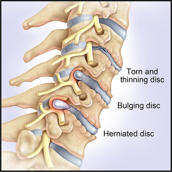
Dr. Soldevilla typically performs this surgery for patients who have cervical disc herniation or cervical degenerative disc disease that causes chronic neck and/or arm pain that hasn’t been relieved from non-surgical treatments.
Through this type of surgery, you:
- Maintain normal motion in the neck
- Have no complication or issues sometimes associated with a bone graft or fusion
- Have a reduced chance of degenerative disc disease in adjacent cervical joints
The procedure is designed to relieve the pressure on your spinal cord or nerves that are causing the pain, numbness, and weakness that can radiate to your shoulder, arm, and hand.
The procedure
Dr. Soldevilla performs the Mobi-C total disc replacement through a small incision on the neck. After he removes the damaged disc, he will smooth away any bone spurs and then place and anchor the Mobi-C artificial disc in the empty space that is left. Dr. Soldevilla then closes the incision and covers it with a dressing.
You will then be moved to a recovery room where you will be monitored for the next few hours. After you have recovered, you will be sent home with post-op instructions. Dr. Soldevilla may also recommend you wear a soft collar for a short period of time to add support to your neck.
Anterior Cervical
Discectomy Fusion
(ACDF)
What is it?
Dr. Soldevilla performs this surgery to remove a damaged disc caused by a cervical herniated disc, cervical degenerative disc disease, or cervical spinal stenosis. The procedure is designed to relieve pressure on the spinal cord or nerve root and alleviate the associated pain and symptoms such as:
- Neck pain
- Radiating pain in shoulders, arm, and hand
- Numbness/tingling in the arm and hand
- Weakness in arm and hand
- Headaches
- Stiffness and difficulty turning your head
The procedure
Dr. Soldevilla performs this surgery using a minimally invasive technique where he makes a small incision on the front (anterior) of the neck, which gives him direct access to the disc through a relatively uncomplicated pathway.
The surgery itself is made up of two parts:
1. Discectomy – First, Dr. Soldevilla makes an incision through the front of the neck and removes the damaged disc.
2. Fusion – He then inserts a bone graft or an implant device in the empty space where the damaged disc was to provide strength and stability to the area.
The procedure may also involve removing part of the vertebra or widening the spinal canal to give the spinal cord more space if neurological symptoms from spinal cord compression are present.
Because this is a minimally invasive technique, Dr. Soldevilla often performs the procedure on an outpatient basis. Once the procedure is completed, you will be moved to a recovery room and you will be monitored for a few hours. Your recovery should be faster and you should also experience less postoperative pain.


Cervical
Decompression
What is it?
Dr. Soldevilla performs cervical decompression surgery to relieve compression of one or more of the vertebrae located in your neck. This compression can occur for a number of different reasons. One of the most common is the gradual wear and tear on the cervical spine that typically occurs in people over 50 years of age.
Other conditions that cause cervical compression usually develop more quickly and occur at any age. These include:
- Injury to the cervical spine
- Scoliosis (abnormal spine alignment)
- Bone disease
- Spinal tumor
- Certain bone diseases
- Rheumatoid arthritis
- Infection
Symptoms of cervical compression include numbness, pain, and weakness. The first line of treatment may involve non-steroidal, anti-inflammatory drugs (NSAIDs) for relief of pain and swelling; physical therapy to strengthen your neck muscles, bracing to provide support; and/or steroid injections. Except in emergency cases, surgery is typically the last resort.
The procedure
Cervical decompression surgery is a procedure designed to remove structures such as bone spurs that are compressing the nerves in your neck while widening the space between vertebrae. Removal of these small structures should alleviate pressure and allow the nerve to heal.
The goal of cervical decompression surgery is to leave the cervical spine intact. However, in some situations, there are so many bony structures pressing on the nerve that removing them may cause instability. If this is the case, cervical decompression surgery Dr. Soldevilla may also perform cervical fusion surgery to correct the instability.

Posterior
Cervical
Fusion
What is it?
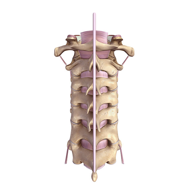
Posterior cervical fusion is the general term used to describe the procedure of mending two or more cervical vertebrae through an incision in the back of the neck.
This procedure is a common one for patients who have cervical spine fractures or instability. A posterior cervical fusion may also be recommended to straighten the cervical spine and stop the progression of a spinal deformity, or to remove a tumor(s).
The goal of the procedure is to relieve pressure on the spinal cord and nerves, or to provide neck stability by fusing two or more vertebrae to into one solid bone.
The procedure
During a posterior cervical fusion, Dr. Soldevilla makes an incision in the back of the neck and places a bone graft along the sides of the injured section of the cervical spine. Over time, this bone graft fuses together to provide healing and greater stability.
Metal screws and rods are sometimes used in conjunction with the procedure to extend fusion and/or provide immediate stability and increase the likelihood of successful fusion.

