Back

What is it?
This minimally invasive procedure is performed to relieve pressure on the spinal nerve column caused by a herniated lumbar (lower back) disc. The goal of the procedure is to decrease pain and allow you to regain normal movement and function.
Dr. Soldevilla performs a microdiscectomy using a special microscope to view the disc and nerves. Using this microscope gives Dr. Soldevilla a larger view, which, in turn, allows him to make a smaller incision. The smaller the incision, the less damage is made to surrounding tissue.
The procedure
The goal of a microdiscectomy is to remove the disc material placing pressure on the nerves by the herniated disc. Since the procedure is done under general anesthesia, you’ll be unconscious during the entire procedure.
With the patient lying face down, Dr. Soldevilla will make an incision directly over the affected disc. Dr. Soldevilla will then remove the damaged tissue and close the incision with sutures. Patients are typically discharged the same day or the morning after an overnight stay in the hospital.
Lumbar
Laminectomy
What is it?
A lumbar laminectomy is a type of back surgery that enlarges your spinal canal in the lumbar area (lower back) by removing the lamina – the back part of a vertebra covering your spinal canal. Enlarging your spinal canal relieves pressure on the spinal cord or nerves caused by bone spurs within the spinal canal.
These bone spurs can narrow the space available around your spinal cord and nerves and can cause pain, weakness, or numbness that radiates down your arms or legs.
The procedure
During a lumbar laminectomy, you will be under anesthesia and won’t feel anything. Dr. Soldevilla will then make a small incision in your back directly over the affected area and remove part or all of the lamina bones as well as any bone spurs or small disc fragments.
Depending on your condition, Dr. Soldevilla may also need to perform a spinal fusion of two or more vertebrae to better stabilize the spine, or a foraminotomy to widen the area where the nerve roots go through the spine.


Fusion
(TLIF, PLIF,
& XLIF)
What is it?
Dr. Soldevilla performs a minimally invasive spinal fusion to permanently “fuse” two or more vertebrae together in your spine in order to eliminate motion between them.
During the procedure, Dr. Soldevilla makes a small incision and removes the damaged disc. He then places a bone graft spacer between two vertebrae to restore height and relieve nerve pinching. As your body begins to heal, new bone grows around the graft, fusing the two vertebrae into one solid piece of bone in about three to six months.
Spinal fusion is used to treat:
- Spinal weakness or insecurity due to a condition such as severe arthritis of their spines. The condition has caused excessive motion between the joints and spinal fusion restores stability.
- Herniated discs – When these are removed, your spine can destabilize. Fusing the vertebrae can provide stabilization.
- Spine deformities – Fusion can help correct spine deformities like scoliosis.
The procedure
There are a number of different ways to reach the spine and perform spinal fusion. Which one Dr. Soldevilla will use depends on the location of your damaged disc and your own unique needs.
TLIF
TLIF (Transforaminal Lumbar Interbody Fusion)
During this procedure, Dr. Soldevilla makes the incision off the middle of your lower back. He then removes the facet joint in order to enlarge the foramen opening as well as any bone spurs and/or ligaments that are compressing the nerve. He then removes the damaged disc and fills the space with a bone graft spacer, which is strengthened with metal screws and rods. TLIF fuses both the front disc space and the back facet joints, stopping all motion at that level of the spine.
PLIF
PLIF (Posterior Lumbar Interbody Fusion)
As with the TLIF, the goal of a PLIF is to fuse vertebrae together to eliminate movement. One of the differences between the procedures is that PLIF achieves spinal fusion by inserting a cage of either gone graft or synthetic material (like titanium) directly into the disc space left by the removal of the damaged disc and bone grows around it.
XLIF
XLIF (eXtreme Lateral Interbody Fusion)
The XLIF procedure differs from the other two procedures in that the spine is accessed through an incision on the side above the hip bone rather than from the front or back.
Sacroiliac (SI)
Joint Fusion
What is it?
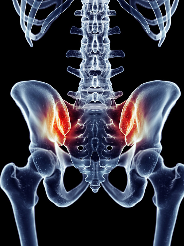
Your sacroiliac joint (also known as your SI joint) connects your hip bones to either side of your sacrum (tail bone), acting as a sort of shock absorber for your lower body and torso. Sacroiliac joint fusion surgery is typically performed when:
- You have significant pain in your lower back, hip, or groin that makes it difficult to function in everyday life
- Nonsurgical methods have not been effective
- There is instability in your pelvis and lower back that causes pain or makes it difficult to stand or walk
- You have stiffness and limited mobility in your hips, lower back, groin, or legs
- There is increased pain after sitting, standing, or lying for long periods of time
The procedure
Almost all of the sacroiliac joint fusion surgeries that Dr. Soldevilla performs as minimally invasive procedures using the most advanced techniques and tools.
After making small incisions on your hip, Dr. Soldevilla uses X-ray scans to view where instrumentation needs to go. He will then drill holes in the sacrum and ilium where he places implants to make the joint more stable. Dr. Soldevilla may also place a bone graft to the ilium to encourage bone growth across the joint. Fusion occurs during the healing process following the surgery.
The procedure usually takes about an hour and you will most likely be in the hospital overnight. Afterward, you will walk on crutches for a few weeks, and then we will allow increased activity.


